Dental Planning Lab
  • Home
  • Services
  • Workflow
  • Pricing
  • About
  • Blogs
  • Contact
Submit a Trial Case
  • Home
  • Services
  • Workflow
  • Pricing
  • About
  • Blogs
  • Contact
  • Submit a Trial Case

Dental Planning Lab

Your Digital Dental Design Partner.

Quick Links

  • Home
  • Services
  • Workflow
  • Pricing
  • About
  • Blogs
  • Contact

Services

  • Aligner Treatment Planning
  • Implant Planning
  • Complete Denture Design
  • Partial Denture Design
  • Restorative Dentistry

Get in Touch

  • info@dentalplanninglab.com
  • +92 310 9076633
  • Lahore, Pakistan
  • Mon – Sat

© 2026 Dental Planning Lab. All rights reserved.

Made by Hamza Ejaz

Privacy PolicyTerms of Service
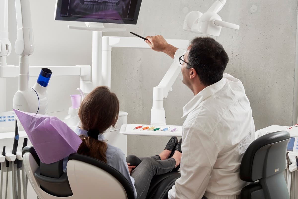
Clinician analyzing a panoramic dental X-ray on a monitor for implant planning
Implant Planning·5 min read·August 14, 2024·By Dental Planning Lab Team

CBCT to Implant Plan: The Complete Digital Workflow

CBCT imaging transformed implant planning from two-dimensional guesswork into three-dimensional precision, but the value of a CBCT scan depends entirely on how clinicians and planners process that data into an actionable treatment plan. This article traces the complete pathway from scan acquisition through implant plan delivery, covering the technical steps our team executes when processing cases submitted via our digital workflow.

Clinical Benefits

  • Three-dimensional visualization of bone anatomy, nerve pathways, and sinus boundaries
  • Accurate implant sizing and positioning based on volumetric bone assessment rather than periapical estimates
  • Seamless transfer from planning data to surgical guide fabrication without manual remeasuring
  • Documented planning records that support informed consent and medicolegal documentation

Clinical Applications

From routine cases to complex multidisciplinary treatment, the following applications are where digital planning delivers the most value for clinics, laboratories, and specialists.

  • Posterior mandible cases requiring inferior alveolar nerve identification and safety zone planning
  • Maxillary implant sites near the sinus floor where grafting or lift planning is needed
  • Multi-implant cases where parallelism and distribution must be verified in three dimensions
  • Immediate placement scenarios where extraction socket morphology affects implant trajectory

Digital Workflow

A predictable digital workflow reduces remakes, shortens chair time, and improves communication between the clinic and planning lab.

  1. Acquire CBCT with appropriate field of view and resolution for the planned implant sites
  2. Segment anatomy: mandibular canal, maxillary sinus, cortical plates, and relevant pathology
  3. Merge intraoral scan or model scan with CBCT using radiographic stent or matching landmarks
  4. Position implants with prosthetic overlay and verify safety margins to critical structures
  5. Design and export surgical guide for printing or milling, with drilling protocol documentation

Clinician analyzing a panoramic dental X-ray on a monitor for implant planning
Digital planning connects clinical records with lab-ready design outputs.

Best Practices

Planning tip

Submit complete records early—photos, scans, and bite data—so planners can flag risks before design begins.

  • Use a limited field of view CBCT centered on the implant site for optimal resolution
  • Apply radiographic stents with fiduciary markers when merging CBCT with intraoral scans
  • Maintain a minimum 2 mm safety margin from the inferior alveolar nerve unless documented otherwise
  • Review merged data for alignment errors before committing to guide fabrication

Common Mistakes to Avoid

  • Using panoramic or periapical radiographs alone for implant depth planning in complex anatomy
  • Failing to verify CBCT-to-scan merge accuracy, resulting in guide misalignment at surgery
  • Ignoring artifact from existing metal restorations that obscure bone assessment in CBCT
  • Selecting implant length without accounting for apical bone concavity visible only in CBCT

“Accuracy in planning is not about more software—it is about better inputs, experienced review, and manufacturing-aware design decisions.”

— Dental Planning Lab clinical team

Conclusion

Strong outcomes in cbct to implant plan: the complete digital workflow depend on clear clinical goals, accurate records, and a planning partner who understands manufacturing requirements. Explore our specialist service, review the case submission workflow, or contact our team to discuss your next case.

Key Takeaways

  • CBCT is the foundation of modern implant planning but requires expert segmentation and merging
  • Prosthetic overlay on CBCT data bridges surgical and restorative planning in one model
  • Surgical guides are only as accurate as the data merge and planning decisions upstream
  • Outsourced planning teams process CBCT data daily, reducing errors common in occasional planners

Table of Contents

  1. Clinical Benefits
  2. Clinical Applications
  3. Digital Workflow
  4. Best Practices
  5. Common Mistakes to Avoid
  6. Conclusion

FAQ

Frequently Asked Questions

Use a voxel size of 0.2 to 0.3 mm with a field of view that includes the planned implant sites plus adjacent anatomical landmarks. Small field of view scans centered on the site deliver better resolution than full-head scans for single or regional implant planning.

Planners use radiographic stents with gutta percha or metal markers visible in both CBCT and optical scan, or match anatomical landmarks such as cusps and incisal edges. Software algorithms refine the alignment, which planners verify visually before implant positioning.

Guides are strongly recommended for multiple adjacent implants, angulated implants, sites near critical anatomy, and full arch cases. Single posterior implants in abundant bone may be placed freehand with a detailed planning report, though guides still improve accuracy.

Standard cases typically return planning proposals within three to five business days after complete records are received. Complex full arch cases with provisional design may require additional review cycles. Guide fabrication adds one to three days depending on printing method.

Keep Reading

Related Articles

Cross-section dental implant model showing prosthetic crown and bone integration
Implant Planning·5 min read·July 8, 2024

Prosthetically Driven Implant Planning: Step-by-Step Guide

Prosthetically driven implant planning starts with the final restoration in mind. Follow this step-by-step guide to position implants for optimal aesthetics, function, and long-term success.

Read Article →
Patient fitting a clear aligner tray during orthodontic treatment planning
Aligner Treatment Planning·5 min read·January 15, 2024

Clear Aligner Treatment Planning: A Complete Guide for Dentists

Master the fundamentals of digital clear aligner treatment planning—from diagnosis and staging to attachments, refinements, and lab collaboration—for predictable orthodontic outcomes.

Read Article →
Dentist reviewing a panoramic dental X-ray with a patient during case consultation
Aligner Treatment Planning·5 min read·June 22, 2024

How to Submit Your First Aligner Case to a Planning Lab

A step-by-step walkthrough for submitting your first clear aligner case to a planning lab—records, prescriptions, file formats, and what to expect during review and approval.

Read Article →

Ready to Discuss Your Next Digital Dental Case?

Partner with Dental Planning Lab for accurate planning, expert digital design, and manufacturing-ready outputs trusted by dental professionals worldwide.

Submit a Trial CaseContact Us